Why Traditional X-Rays Miss Critical Details
Standard dental X-rays have served dentistry for over a century, but they come with a fundamental limitation: they flatten three-dimensional structures into two-dimensional images. When a dentist needs to evaluate bone density, locate hidden infections, or plan implant placement, flat images leave too many questions unanswered. That's where cone beam computed tomography (CBCT) changes the game.
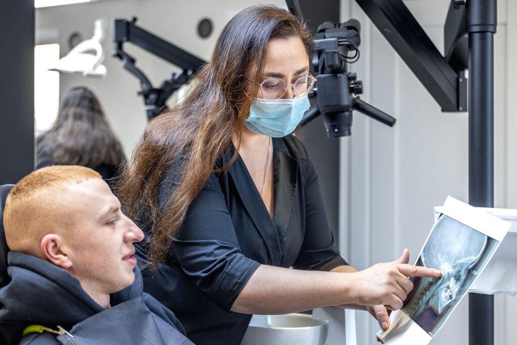
Dr. Ryan Watkins, founder of Dreamtime Dentistry in Vista, California, has built his practice around advanced imaging technology. As a dentist anesthesiologist with training from Loma Linda University, he understands the stakes when treatment planning goes wrong. His practice uses CBCT scans daily to catch problems that conventional imaging would miss entirely.
"We had a patient come in for what looked like a simple crown. The scan showed a crack running deeper than expected. Without that, we would've treated the wrong problem first," he explains. That single scan prevented a failed restoration and additional procedures down the line.
What CBCT Actually Shows
CBCT scanners create detailed three-dimensional images of teeth, soft tissues, nerve pathways, and bone structure in a single rotation around the patient's head. The technology captures hundreds of images in seconds, then reconstructs them into a 3D model that dentists can manipulate and measure from any angle.
The difference in information quality is dramatic. Traditional X-rays might show bone loss, but CBCT reveals exactly where the loss occurred, how severe it is, and whether adjacent structures are affected. For implant cases, CBCT scans map the exact location of sinuses, nerves, and blood vessels that must be avoided during surgery.
According to research published in the Journal of the American Dental Association, CBCT imaging improves diagnostic accuracy by up to 40% in endodontic cases compared to conventional radiography. In implant dentistry, studies show CBCT reduces surgical complications by identifying anatomical variations before the procedure begins.
Real World Applications in Complex Cases
Dreamtime Dentistry integrates CBCT into workflows for cases where precision matters most. Watkins notes that advanced imaging allows the practice to see more, measure more, and plan with far greater accuracy before treatment begins.
Root Canal Diagnosis
Tooth anatomy varies more than most patients realize. Some molars have four root canals instead of three. Others have canals that curve sharply or split unexpectedly. Missing a canal during root canal treatment is one of the leading causes of procedure failure.
CBCT scans reveal every canal, showing their exact location, curvature, and length. Endodontists can spot calcified canals, fractured roots, and periapical lesions that appear normal on standard films. This information transforms treatment from educated guesswork into precise, measured intervention.
Dental Implant Planning
Placing dental implants requires knowing exactly what lies beneath the gum tissue. Bone density, width, and height determine whether an implant will integrate successfully. Proximity to nerves determines whether the patient will experience numbness or pain after surgery.
With CBCT, dentists measure available bone to the tenth of a millimeter. They can simulate implant placement virtually, adjusting angle and depth before making a single incision. For practices like Dreamtime Dentistry, where Dr. Kyung Boen has over 25 years of experience placing dental implants, CBCT has become as fundamental as the drill itself.
Impacted Teeth and Extractions
Wisdom teeth often grow at odd angles, sometimes wrapping around nerves or pressing against adjacent teeth. Extracting an impacted tooth without knowing its exact position relative to critical structures carries significant risk.
CBCT scans eliminate that uncertainty. Surgeons can visualize the tooth's roots in relation to the inferior alveolar nerve, plan their extraction path, and anticipate complications before they occur. This level of preparation reduces surgical time, minimizes trauma, and improves patient outcomes.
The Diagnostic Workflow Advantage
Watkins emphasizes that CBCT isn't just about seeing more. It's about making better decisions faster. When a patient presents with complex symptoms, pain in an unclear location, or a history of failed treatment, CBCT provides clarity that speeds up diagnosis and builds patient confidence.
The practice uses CBCT imaging to coordinate care across multiple specialists in house. When a patient needs both an implant and a bone graft, the oral surgeon and restorative dentist can review the same 3D scan, plan their procedures in sequence, and anticipate challenges together.
"We didn't want patients going from office to office trying to complete a treatment plan. If we can bring the right expertise into one place, it simplifies everything for the patient," Watkins notes. CBCT plays a central role in making that integrated approach possible.
Radiation Exposure and Safety Considerations
One common concern about CBCT is radiation exposure. While CBCT does expose patients to more radiation than a standard dental X-ray, the dose remains significantly lower than medical CT scans. A typical CBCT scan delivers between 50 and 100 microsieverts of radiation, compared to 400 to 1,000 microsieverts for a medical head CT.
Modern CBCT machines use optimized protocols that reduce exposure while maintaining image quality. Dentists order CBCT scans only when the diagnostic benefit outweighs the minimal risk, not as a routine screening tool.
When CBCT Makes the Difference
Not every dental case requires three-dimensional imaging. Routine cleanings, simple fillings, and straightforward extractions don't need it. But when cases involve infection, trauma, anatomical variation, or surgical planning, CBCT transforms outcomes.
Dr. Ryan Watkins has seen the technology prevent complications, reduce treatment time, and improve patient satisfaction. As practices adopt more advanced tools like intraoral scanners and same-day crown systems, CBCT fills a crucial diagnostic role that makes all other technology more effective.
For patients facing complex dental work, asking whether a practice uses CBCT imaging is a reasonable question. The answer reveals how seriously that practice takes precision, planning, and modern standards of care.