AI Assisted Brain Surgery and the Future of Oral and Maxillofacial Surgery
For years, dentistry has talked about artificial intelligence mainly as a diagnostic tool. AI finds caries on radiographs, flags bone loss, measures anatomy, and helps clinicians interpret images. A recent operation in London suggests that the next chapter may be very different. Instead of analyzing information before treatment, AI may begin assisting surgeons while treatment is actually happening.
At the National Hospital for Neurology and Neurosurgery in London, 48 year old Rhys Hibbert underwent endoscopic removal of an 11 millimeter pituitary tumor that was threatening his vision. During the operation, an AI system analyzed the live endoscopic video and highlighted critical anatomy in real time. The surgeon remained fully in control. The software did not cut tissue, move an instrument, or make autonomous decisions. Its role was closer to a digital second set of eyes, continuously identifying structures the surgeon needed to recognize and avoid.
The outcome was dramatic. Hibbert’s tumor was removed, his sight was preserved, and his visual function improved. But the most important scientific point is easy to lose in the excitement. The patient did well while AI was being used. That does not yet prove he did well because of the AI. This was the first patient in an ongoing clinical trial, not proof that AI reduces complications or improves outcomes. The milestone is real, but it is still primarily a technical milestone.
The work did not appear overnight. In 2024, the PitSurgRT system was trained on video from 64 pituitary operations to identify the sella, carotid arteries, optic protuberances, and other critical anatomy. After optimization, it processed images at roughly 298 frames per second, fast enough for live video. Neurosurgeons judged about 89 percent of landmark predictions sufficiently accurate for guidance. Yet estimated localization error remained roughly 2.35 to 3.10 millimeters. Fast is not the same as safe.
Another 2024 study tested whether AI could improve human recognition of pituitary anatomy. Twenty four participants, from medical students to expert pituitary surgeons, examined operative images with and without AI assistance. Overall recognition improved, but the largest gains occurred among the least experienced participants. Experts improved very little because they were already good at the task. That may point to one of the most practical early uses of surgical AI. Its greatest value may not be making the best surgeon dramatically better. It may be raising the floor, reducing variation, and helping less experienced operators recognize anatomy more reliably.
There was also a warning. The same inexperienced users who benefited most were more likely to change their answers when the AI suggested something different. Sometimes that made them better. Sometimes it made them worse. That is automation bias. The dangerous AI error is not the obvious one everyone rejects. It is the confident, plausible error that appears after the clinician has learned to trust the system.
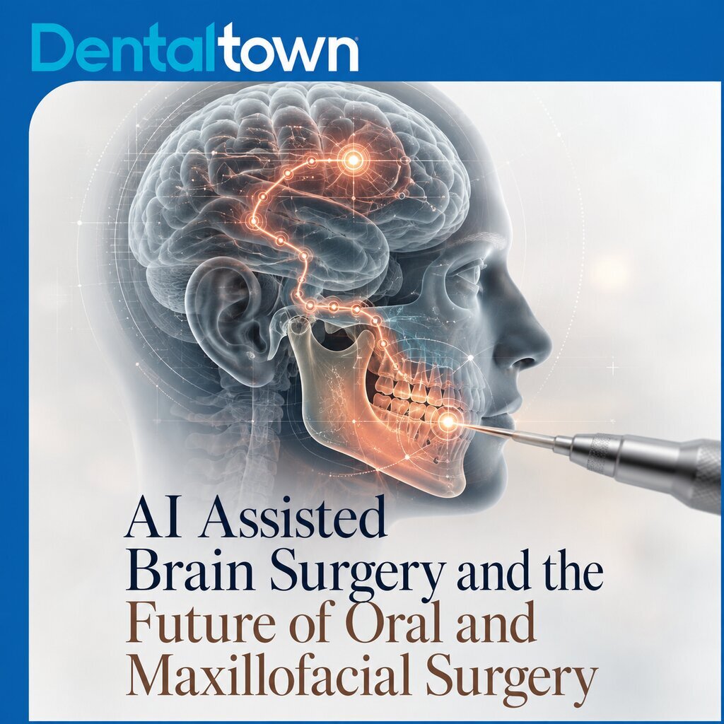
For oral and maxillofacial surgery, the attraction is obvious. Dentistry already has CBCT, virtual surgical planning, static guides, dynamic navigation, augmented reality, robotics, and increasingly sophisticated computer vision. Those technologies are beginning to converge. A future surgeon might see planned anatomy registered directly to the patient while software recognizes the operative field and tracks the bur or saw. The system could potentially highlight the expected location of the inferior alveolar canal, mental foramen, maxillary sinus, osteotomy line, tumor margin, or vascular structure and warn when an instrument approaches a danger zone.
Oral and maxillofacial surgery is already one of the most active specialties in augmented reality research. A 2026 systematic review of clinical head mounted navigation systems included 61 studies involving 1,547 patients, with oral and maxillofacial surgery contributing more studies than any other specialty. The technology has been investigated in orthognathic surgery, trauma, orbital reconstruction, tumor surgery, implant placement, and craniofacial reconstruction.
The accuracy numbers explain why caution still matters. Across 27 studies reporting positional accuracy, the weighted mean error was 2.58 millimeters, with results ranging from 0.8 to 13 millimeters. In a large reconstructive procedure, a few millimeters may be acceptable. Beside an inferior alveolar nerve, tooth root, optic nerve, or artery, it may not be. A beautiful hologram is clinically useless if it is three millimeters away from the anatomy it claims to represent.
That may be the central technical challenge for dentistry. The London system primarily interprets live endoscopic video. An oral surgery system would often need to do more. It would have to recognize the live field, track the instrument, and keep a preoperative CBCT accurately registered to anatomy that may move, deform, bleed, or become obscured by irrigation, suction, retractors, and tissue changes. The registration problem may prove harder than the AI itself.
Workflow and economics will matter just as much as engineering. A sophisticated navigation system could make enormous sense in oncologic resections, orbital reconstruction, orthognathic surgery, craniofacial reconstruction, or zygomatic implant placement. It may make far less sense for a routine extraction if setup, calibration, disposables, training, and equipment cost outweigh the incremental benefit. Adoption will likely begin where anatomy is difficult, consequences are high, and precision has real clinical and financial value.
The winning interface may not even be an augmented reality headset. Today’s headsets remain heavy, imperfect, difficult to sterilize, and prone to visual fatigue and registration drift. The same information could eventually appear through a surgical microscope, endoscope, loupes, robotic console, or navigation display already familiar to the operator.
For practicing dentists, the useful framework is simple. CBCT shows where anatomy should be. Navigation shows where the instrument is. Computer vision recognizes what the surgeon is seeing. AI may eventually combine those streams and warn when something important is about to happen.
That is a much bigger leap than finding caries on a bitewing. It moves AI from interpretation before treatment to assistance during treatment. The promise is compelling, but the evidence remains immature. In the 2026 augmented reality review, most studies were rated as low or very low certainty, and only two were randomized trials. Before this becomes routine care, dentistry will need multicenter validation, better registration, lower error rates, reliable failure detection, and proof that the technology improves patient outcomes rather than simply making the operatory look more advanced.
The London case matters because real time surgical AI has now crossed from research videos into a live human operation. What it does not yet show is that AI reliably makes surgery safer. That distinction should shape how dentistry watches the next wave of innovation.
When the digital second set of eyes arrives in your operatory, will it make you a better surgeon, or simply give you one more thing to trust?
Join the Conversation!
Sources
First live AI assisted brain surgery
First patient in live AI assisted sight saving brain surgery. University College London, 2026. https://www.ucl.ac.uk/news/2026/aug/first-patient-live-ai-assisted-sight-saving-brain-surgery
Computer Vision for Real Time Anatomical Navigation in Neurosurgery: First in Human Clinical Evaluation and Iterative Development. medRxiv, 2026. Preprint. https://www.medrxiv.org/content/10.64898/2026.06.11.26355205v1
Development of real time surgical AI
PitSurgRT: Real Time Localization of Critical Anatomical Structures in Endoscopic Pituitary Surgery. International Journal of Computer Assisted Radiology and Surgery, 2024. https://link.springer.com/article/10.1007/s11548-024-03094-2
Artificial Intelligence Assisted Operative Anatomy Recognition in Endoscopic Pituitary Surgery. npj Digital Medicine, 2024. https://www.nature.com/articles/s41746-024-01273-8
Oral and maxillofacial surgery
From Static to Robotic: Evolving Navigation Systems in Oral and Maxillofacial Surgery. Oral Surgery, Oral Medicine, Oral Pathology and Oral Radiology, 2026. https://www.sciencedirect.com/science/article/pii/S2212440325012799
Clinical accuracy, limitations, and evidence quality
Augmented Reality in Navigated Surgery: A Systematic Review of Clinical Accuracy and System Performance. Mayo Clinic Proceedings: Digital Health, 2026. https://www.mcpdigitalhealth.org/article/S2949-7612(26)00024-6/fulltext